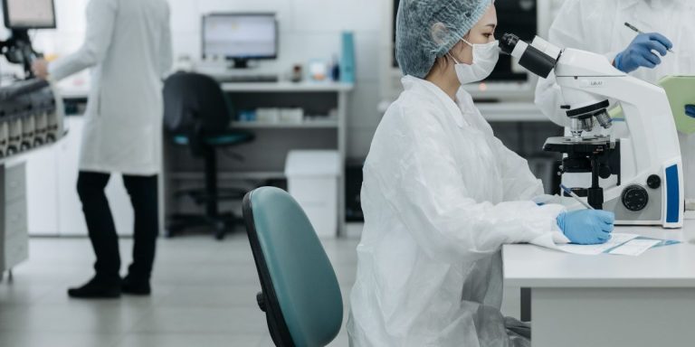
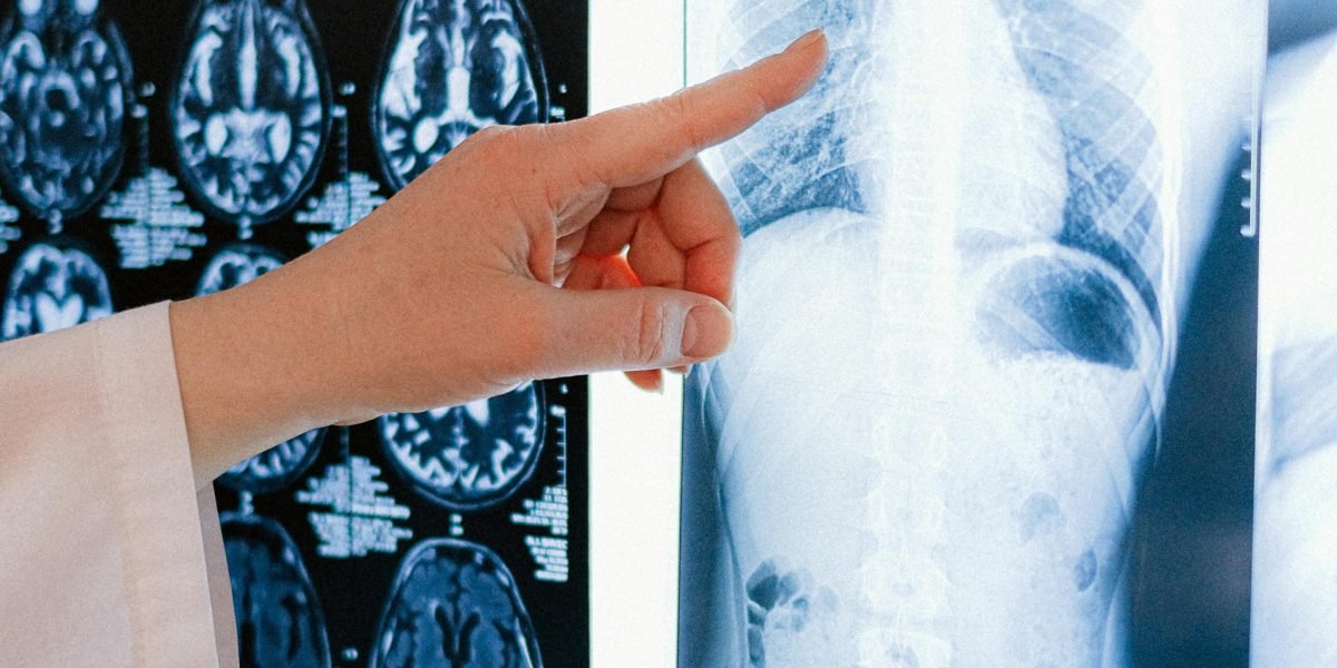
Introduction
Have you ever waited longer than expected for a scan result or worried that a small detail might get missed?
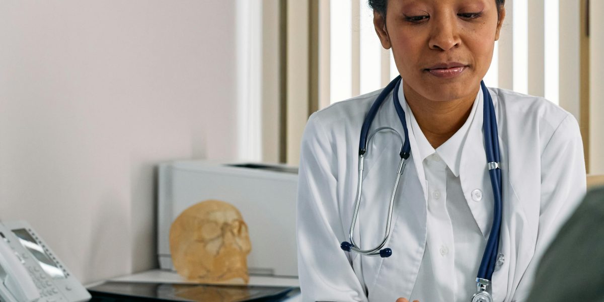
You are not alone. Across the globe, hospitals are struggling with a shortage of radiologists, and diagnostic errors remain a serious concern. In fact, many imaging departments are stretched thin, which can delay care and lead to mistakes. But there is good news. Doctor AI is stepping in to help.
Artificial intelligence with images is no longer just a futuristic idea. In 2026, it is already making a real difference in clinics and hospitals. Right now, about 81% of radiology departments report using some form of artificial intelligence imaging tool, especially for stroke detection and report writing (source: PubMed study).
That is a huge leap forward.
The numbers back up this shift. The AI in medical imaging market is estimated at $2.20 billion in 2026 and is expected to grow fast, hitting $17.77 billion by 2033 (source: Coherent Market Insights). That growth tells us that hospitals and doctors see real value in using an artificial intelligence detector to catch what the human eye might miss.
So what does this mean for you? Whether you are a healthcare professional, a tech enthusiast, or someone who simply wants to understand how machines help doctors make better calls, this article is for you. We will take a close, data-driven look at how doctor AI works in diagnostic imaging today. We will look at real tools, real results, and what is coming next. If you want to see how AI is changing imaging in business too, check out our piece on how artificial intelligence with images is transforming business in 2026.
Let us dive in and see how this technology is saving time, cutting errors, and making healthcare smarter.
The State of AI in Medical Imaging: Adoption, Market Growth, and Impact (2024–2026)
So how fast is doctor AI really spreading through hospitals and clinics? The short answer is: very fast.
Back in 2022, only about 10% to 15% of radiology practices in the U.S. and Europe had adopted any AI tool. By 2026, that number has more than tripled. A recent study found that over 40% of practices now use at least one artificial intelligence imaging tool, and some reports put that figure even higher for larger hospital systems. In fact, a survey published on PubMed shows that 81% of radiology departments currently report using some form of AI, especially for detecting strokes and helping with report writing (source: PubMed study). That is a huge jump in just a few years.
The money flowing into this space tells the same story. The global AI in medical imaging market is estimated at $2.20 billion in 2026 and is expected to grow at a compound annual growth rate (CAGR) of about 34.8%, reaching $17.77 billion by 2033 (source: Coherent Market Insights).
Another report by Research and Markets values the market at $5.29 billion in 2026 and projects it to hit $20.28 billion by 2030, growing at a 39.9% CAGR (source: Research and Markets). Different numbers, same direction: rapid growth.
What is driving this explosion? Three big reasons stand out.
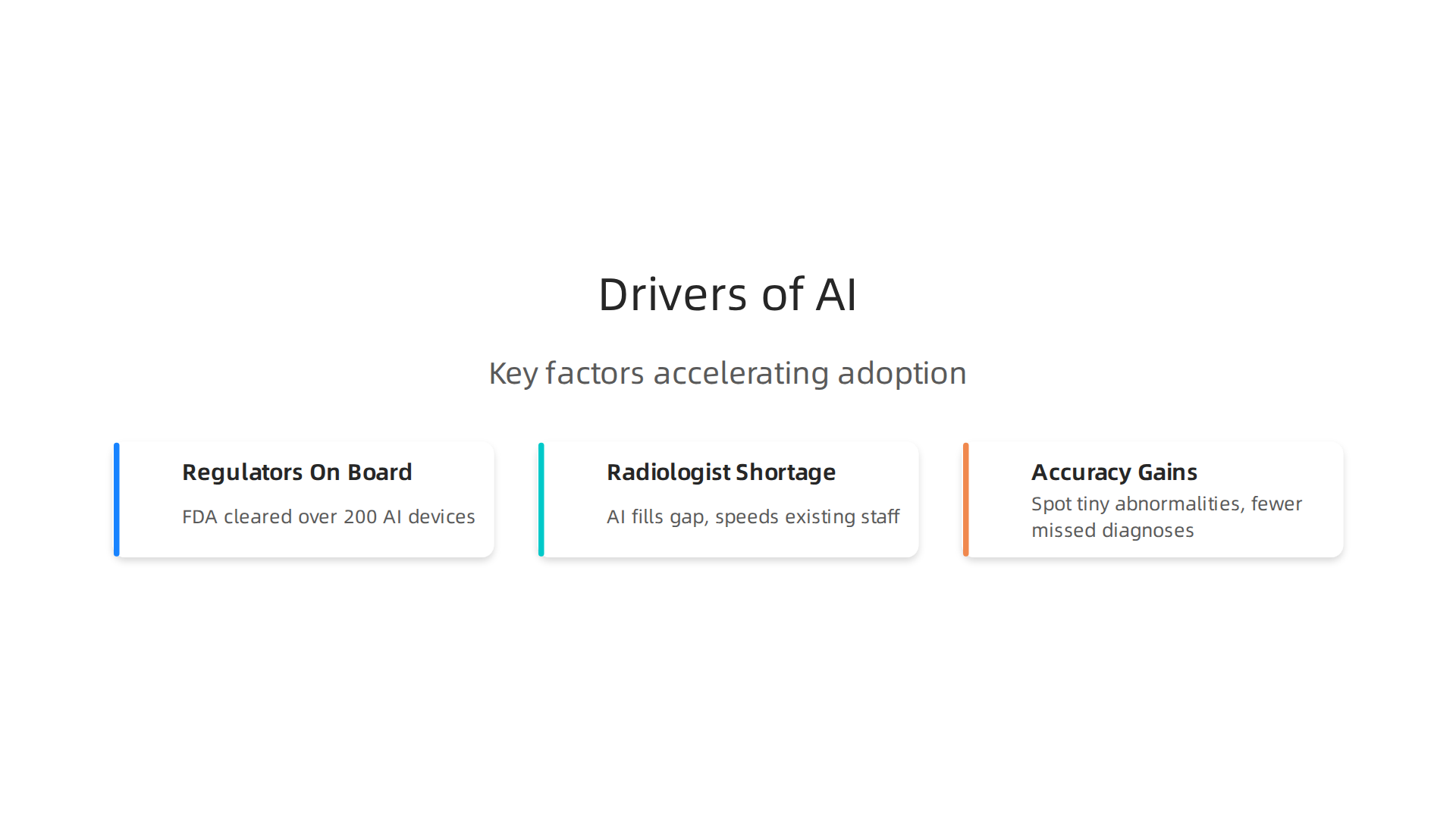
First, regulators are on board. By 2026, the FDA has cleared more than 200 AI-powered medical devices. That gives hospitals confidence to buy and deploy an artificial intelligence detector without worrying about legal hurdles. Second, there is a serious shortage of radiologists. Many imaging centers simply cannot hire enough human experts to read all the scans. Doctor AI helps fill that gap, letting existing staff work faster and focus on complex cases. Third, the accuracy gains are real. AI tools can spot tiny abnormalities in CT scans, MRIs, and X-rays that even trained eyes might miss. That means fewer missed diagnoses and better patient outcomes.
For a deeper look at how this same technology is changing business, check out our article on how artificial intelligence with images is transforming business in 2026.
The bottom line: artificial intelligence imaging is no longer experimental. It is a standard tool that is growing fast, backed by strong market data and real clinical results. As we move through 2026, expect even more hospitals to bring in doctor AI to help with everything from detecting lung nodules to spotting early signs of cancer. The future of diagnostics is here, and it is powered by smart algorithms working side by side with human doctors.
Core AI Technologies Powering Diagnostic Applications
So what actually makes doctor ai work behind the scenes? The engine is deep learning, and the workhorse is the convolutional neural network (CNN).
CNNs are really good at finding patterns in images. In fact, they dominate the medical imaging AI market with a 48% share Precedence Research.
But they aren’t the only player anymore.
A newer approach called transformers is now competing with CNNs. These models use attention mechanisms to understand the whole image at once. Researchers are building hybrid CNN-transformer models that combine the strengths of both. A recent study showed one such model successfully distinguishing between different types of breast lesions with high accuracy SAGE Journals.
Another big challenge is data. Artificial intelligence imaging needs lots of labeled medical images, but those are hard to get because of privacy rules. That is where synthetic data and generative AI come in. These tools create realistic but fake medical images that help train artificial intelligence detectors without using real patient data. For a look at how similar image generation tech is changing business, read our guide on AI image generation in 2026.
Together, these technologies are making doctor ai faster, smarter, and safer for everyday use.
Deep Learning and Convolutional Neural Networks (CNNs)
Even with newer models like transformers getting attention, convolutional neural networks remain the backbone of most doctor ai tools. Why? Because CNNs are incredibly efficient at spotting patterns in images. They scan pixel by pixel, looking for edges, shapes, and textures that tell a radiologist something important.
Recent innovations have made them even smarter. Attention-gated CNNs learn which parts of an image matter most, so they don’t waste time on background noise. 3D CNNs work directly on CT and MRI volumes, analyzing slices together instead of one at a time. This helps artificial intelligence imaging tools catch things like tiny tumors that might be missed in a single 2D view.
Benchmark numbers back up the progress. On the big public datasets like CheXpert and MIMIC-CXR, many CNN-based artificial intelligence detectors now hit an AUC of 0.95 or higher for common chest conditions. That means they separate sick from healthy with near-human accuracy.
The latest wave also combines CNNs with other deep learning techniques. One framework now integrates radiology and pathology imaging into a single model, making artificial intelligence with images even more powerful for complex diagnoses The Pathologist. And researchers are building foundational models that train on enormous datasets, pushing the limits of what doctor ai can do RSNA.
Want to see how artificial intelligence with images is changing medicine? Check out our guide on how artificial intelligence with images is transforming business in 2026.
Natural Language Processing for Radiology Reports
CNNs handle the images. But what about the words radiologists write afterward? Every scan generates a free text report, and those reports hold critical details. Natural language processing, or NLP, is stepping in to automate this side of the workflow.
Here is how it works. NLP models scan the radiologist’s notes and pull out structured findings automatically. Instead of a human reading through pages of text to find key observations, the artificial intelligence detector extracts them in seconds. Large language models like GPT-4 and Med-PaLM 2 now go a step further. They generate preliminary report impressions that radiologists can review and approve.
The results are impressive. Studies from 2025 and 2026 show that using LLMs for this task can cut reporting time by 30 to 50 percent while keeping accuracy high. That means radiologists spend less time typing and more time on patient care. Foundational medical imaging models trained on massive datasets are making these NLP tools even more capable PMC.
This blend of artificial intelligence with images and natural language processing is another reason why doctor ai tools keep getting better. If you want to explore more about how AI is reshaping industries, check out our breakdown of AI image generation market trends in 2026.
Generative AI and Synthetic Data
NLP helps doctors read reports faster. But here is another twist. Generative AI can now create realistic medical images that have never come from a real patient. Sounds a bit like science fiction, right? Actually, it is already happening in 2026.
Generative adversarial networks, or GANs, and diffusion models are the engines behind this. These artificial intelligence detectors learn the patterns in thousands of real scans. Then they produce new, synthetic images that look just as authentic. The goal is not to replace real images. It is to solve a big problem: there are never enough scans to train an AI model well.
Synthetic data helps in two important ways. First, it bulks up small datasets so the artificial intelligence with images learns more reliably. Second, it lets researchers simulate diverse patient populations. That reduces bias, which is a major challenge in healthcare AI today. Foundational deep learning models for medical imaging need huge datasets, and synthetic data fills the gap PMC.
Regulators are starting to take notice. The FDA now accepts synthetic data in certain validation submissions, as long as the simulation follows strict rules. This is a big step forward for doctor ai tools.
If you want to see how artificial intelligence with images is reshaping other fields, check out our article on how artificial intelligence with images is transforming business in 2026.
Top AI Applications in Medical Imaging and Diagnostics
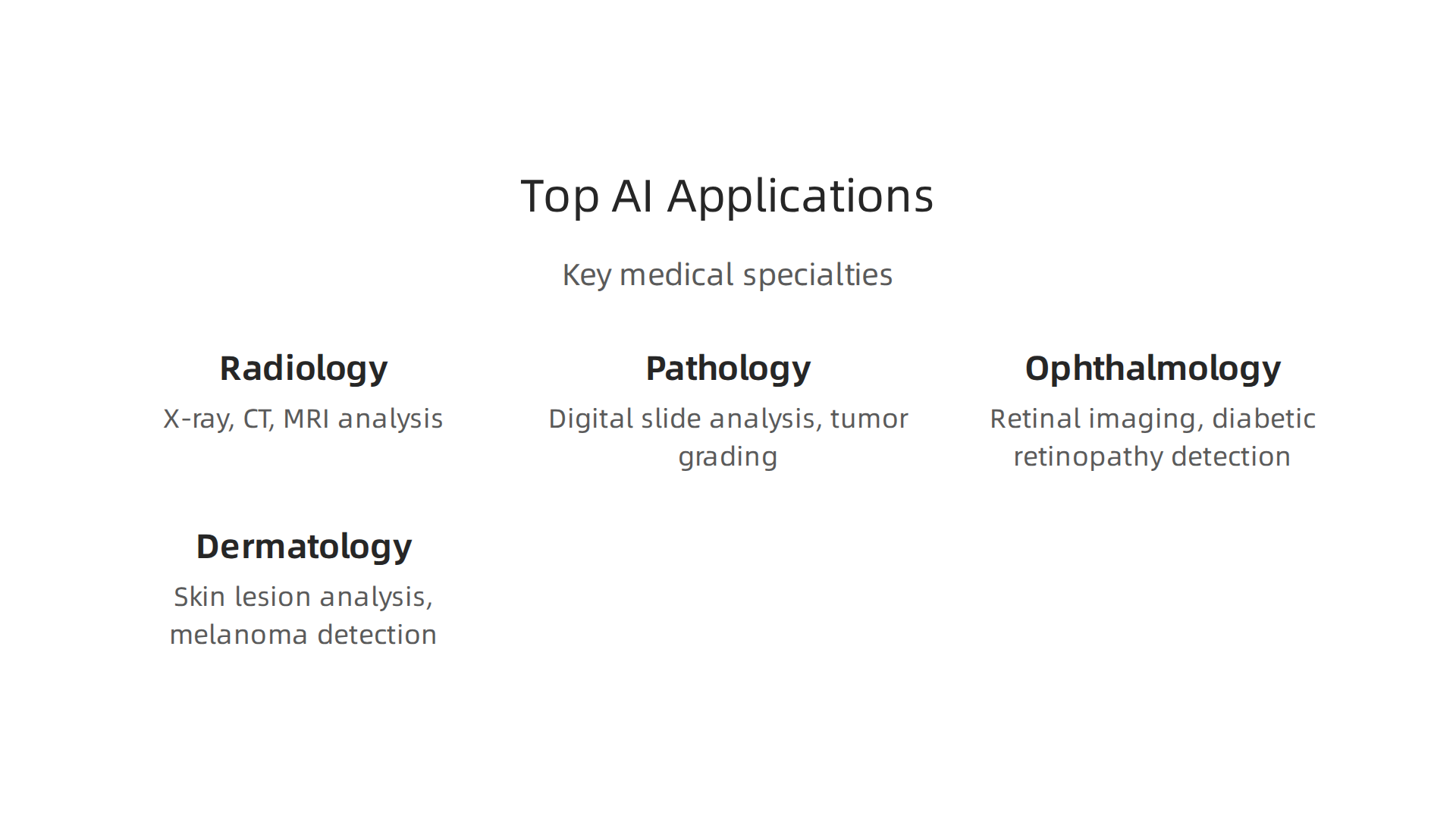
So where does all this actually get used in 2026? It is not just in one department. You will find doctor ai tools working as daily helpers across radiology, pathology, ophthalmology, and dermatology.
Each specialty has a unique workflow. An orthopedic surgeon needs to spot a tiny hairline fracture on an X-ray. A dermatologist needs to check if a mole has changed over time. These are very different tasks, and a smart artificial intelligence imaging tool learns the specifics of each one.
For example, orthopedic AI now acts as a diagnostic assistant. It reviews X-rays using deep learning to help doctors catch fractures they might otherwise miss AZMed. This reduces errors and improves how many patients doctors can see every day.
Radiology leads the pack when it comes to clinical proof. In 2026, radiology AI is less about flashy demos and more about real workflow integration and quality oversight Vestarad. Hospitals now make AI pass strict pilot tests before letting it near patients JMIR.
Ophthalmology is right behind radiology. Eye doctors use artificial intelligence with images to scan retinas for early signs of diabetic blindness or glaucoma. The evidence is strong because the images are standard and the tasks are clear.
This push for hard evidence is healthy. It means every artificial intelligence detector tool has to prove it makes a real difference. As one expert put it, 2026 is the year healthcare AI has to "prove it or move aside" Rad AI.
This doctor ai trend is not just changing hospitals. You can see how how artificial intelligence with images is transforming business in 2026 is reshaping quality and speed across entire industries.
Radiology: X-ray, CT, and MRI
Radiology is where doctor ai tools have gone from experimental to essential in 2026. If you walk into an emergency department today, you will find AI helping with chest X-rays every single day.
Here is what that looks like in practice. AI triage tools now scan chest X-rays for pneumothorax (collapsed lung) and lung nodules automatically. When the artificial intelligence detector finds something urgent, it flags the study immediately. This means the radiologist sees the critical case first, not just the next one in the queue.
CT and MRI have taken this even further. Modern artificial intelligence imaging models now detect stroke, brain hemorrhage, and liver lesions with sensitivity above 95%. That is a huge deal for time sensitive conditions like stroke, where every minute saved can mean less brain damage and a better recovery for the patient.
The real benefit, though, is the workflow gain. Hospitals report that AI cuts radiologist reading time by 20 to 30 percent for normal studies. That might not sound dramatic, but it means doctors spend less time on routine scans and more time on complex, unusual cases that actually need their full attention.
This shift is not just about speed. It is about trust. In 2026, hospitals require AI tools to pass strict pilot tests before they can help with real patients JMIR. The focus is on governance and quality oversight, not just flashy features Vestarad.
If you are curious about how similar artificial intelligence with images tools are changing work across other industries, check out how AI image generation is reshaping business in 2026. It covers ground beyond just healthcare.
Pathology: Digital Slides and AI
Just like in radiology, the doctor ai revolution is reshaping pathology in 2026. Labs are now using digital slide systems that have received real regulatory approval for serious cancers like prostate, breast, and melanoma.
Here is what is different. These AI tools do not just find suspicious spots. They can actually grade tumors. For prostate cancer, studies show that AI matches the accuracy of trained pathologists for tasks like Gleason grading. That is a huge win for standardization and speed.
But 2026 is also the year healthcare AI had to prove its real value. The industry is demanding that these tools prove they work or get out of the way RadAI. So these digital pathology systems act as a careful diagnostic assistant, not a replacement Azmed.
Of course, there are real hurdles. Storing whole-slide images takes massive computing power. Different scanners produce slightly different images, which can confuse the artificial intelligence detector. And some pathologists are hesitant to trust a machine. Hospitals are solving this with strong governance frameworks that test the AI against strict quality standards before it ever helps with a patient Vestarad.
The rise of artificial intelligence imaging in pathology proves one thing: the way we use artificial intelligence with images is getting more mature across every industry. If you want to see how these same visual AI tools are creating value in business and marketing, check out this guide on AI image generation in business.
Ophthalmology: Retinal Imaging and Diabetic Retinopathy
Here is a problem you might know all too well. Diabetic retinopathy is one of the leading causes of blindness, yet many people with diabetes skip their yearly eye exam. That is where doctor ai steps in.
Systems like IDx-DR are now widely deployed in primary care clinics and eye centers. These tools use an artificial intelligence detector to scan retinal images for signs of disease. In clinical studies, they show sensitivity above 90% and specificity above 85% for referable diabetic retinopathy. That means the artificial intelligence imaging catches most cases and rarely flags healthy eyes by mistake.
The approach fits right into the 2026 healthcare trend: AI must prove its real value before it gets used widely RadAI. These retinal systems have been tested against strict standards, just like any other serious diagnostic tool.
But the field is not stopping at diabetic retinopathy. New artificial intelligence with images applications now help detect age-related macular degeneration and glaucoma using optical coherence tomography (OCT) scans. The same visual pattern recognition that works for the eye also powers breakthroughs in other areas. Want to see how these AI imaging tools create value beyond medicine? Check out this guide on AI image generation in business.
Dermatology: Skin Lesion Analysis
From the back of the eye to the surface of the skin, the same doctor ai technology is making a big impact. Algorithms trained on thousands of dermoscopic images can now detect melanoma with accuracy that matches board-certified dermatologists. This is not a future promise. It is happening in clinics right now in 2026.
Consumer apps and teledermatology platforms are adding these artificial intelligence detector tools fast. You can take a photo of a suspicious mole with your phone and get a risk score in seconds. That sounds helpful, but it also raises real concerns. How accurate are these consumer tools in the real world? What happens to your medical images and your privacy? Not every app is held to the same clinical standard.
That is why regulators are stepping in. The FDA now requires real-world evidence before approving skin lesion AI tools for clinical use. This is a major shift. These artificial intelligence imaging systems must prove they work in actual clinics, not just in a controlled lab. The days of releasing an app without solid proof are ending. The entire healthcare AI field is moving toward this "prove it or move aside" standard, where real clinical validation is no longer optional RadAI.
This validation trend is not limited to dermatology. As more industries rely on artificial intelligence with images, the bar for quality and safety keeps rising. If you want to see how this trend plays out beyond the clinic, read our breakdown of AI image generation 2026 market trends and business applications.
Regulatory Landscape: FDA, CE, and Global Standards
All this talk about clinical validation for doctor ai tools in dermatology points to a bigger story. Regulators around the world are rewriting the rulebook for artificial intelligence imaging systems. And in 2026, the pace of change is faster than ever.
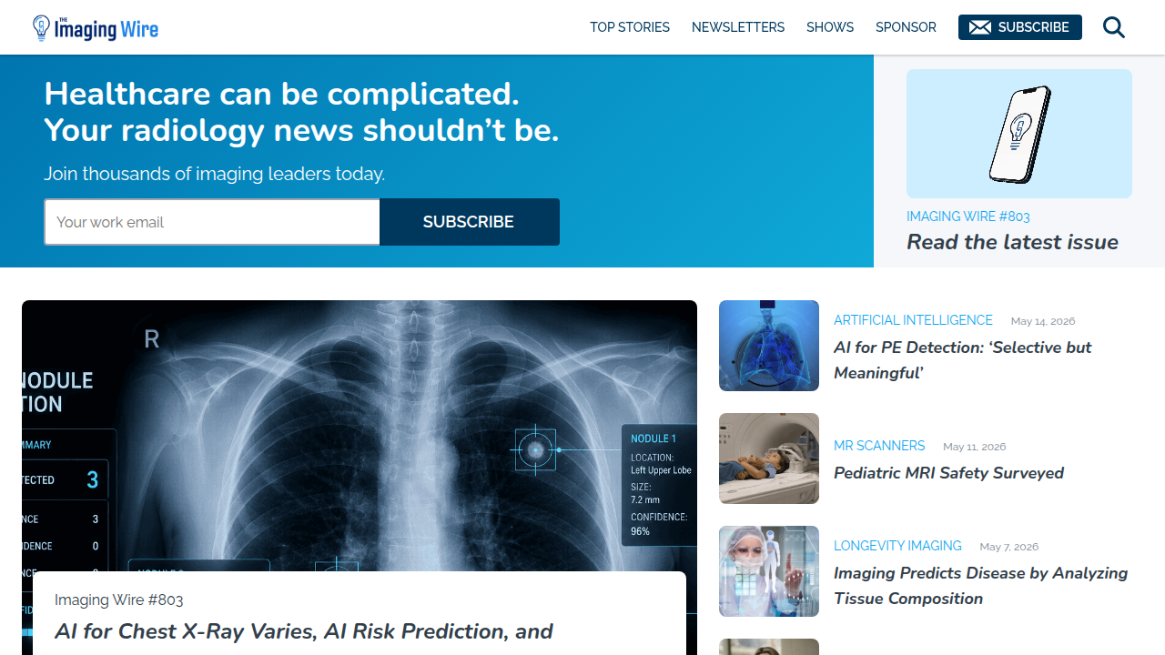
Let us start with the numbers. By early 2026, the FDA had cleared over 200 AI-enabled medical devices for marketing in the United States. Radiology dominates that list by a wide margin. According to the FDA’s updated AI device list, radiology continues to hold the lead as the medical specialty with the most clearances FDA AI Device List.
That makes sense. X-rays, CT scans, and MRIs produce the kind of structured image data that artificial intelligence with images handles really well.
The clearance numbers keep climbing too. Industry reports show a record 295 new clearances in 2025 alone FDA Tracker. But getting approved is only half the battle. The FDA is now asking tougher questions about how these tools perform in real hospitals with real patients, not just in controlled lab settings.
Here is the thing. The old approval system was built for static software. You release version 1.0, get it cleared, and that is it. But AI systems learn and improve over time. That creates a serious problem for regulators. How do you approve a tool that changes every few months?
The FDA answered that question with a new framework called predetermined change control plans, or PCCPs. Instead of requiring a brand new submission every time the AI gets better, manufacturers can describe planned updates in advance. If the updates stay within those pre-approved boundaries, no re-submission is needed Navigating FDA Framework. This is a game changer for artificial intelligence imaging companies that need to ship improvements fast.
Across the Atlantic, the rules are also shifting. Europe’s Medical Device Regulation and In Vitro Diagnostic Regulation are making CE marking harder to get for AI diagnostic tools. The bar for clinical evidence is higher. Companies cannot just run one small study and call it done. They need real-world data that proves their tools work across different patient groups and hospital settings.
The bottom line is this. Whether you are building a skin lesion detector, a chest X-ray reader, or any other doctor ai application, the regulatory path is getting clearer but also harder. The days of releasing an unvalidated tool are over. If you want to stay ahead of these changes, check out our guide on how artificial intelligence with images is transforming business in 2026. It covers the practical steps companies are taking to navigate this new regulatory reality.
Challenges and Limitations: Data Privacy, Bias, and Integration
Even with regulators rewriting the rules for doctor ai tools, big problems still stand in the way. These challenges slow down adoption in real hospitals and clinics. Let’s walk through the three biggest ones.
Algorithmic bias is a serious issue. Many artificial intelligence imaging models are trained on data that does not include enough variety. When the training data comes mostly from one group of people, the model performs poorly on everyone else. For example, a skin lesion detector trained mainly on light skin might miss melanoma on darker skin. This is not just a theoretical concern. Research published in JAMA Network Open found that many FDA-approved AI devices lack strong evidence that they work across different populations Generalizability of FDA-Approved AI Devices. If we want doctor ai to help everyone, the data used to train these tools must reflect real world diversity.
Data privacy regulations create a tough trade off. Laws like HIPAA in the U.S. and GDPR in Europe protect patient information. That is a good thing. But these same rules make it hard to collect the large, diverse datasets needed to fix bias. You cannot just grab medical images from anywhere. You need consent, secure storage, and strict governance. Hospitals often sit on valuable data but cannot share it easily. This tension between privacy and the need for better training data is one of the biggest barriers to building fair and accurate artificial intelligence detector systems.
Integration into clinical workflows is the number one obstacle on the ground. You can have the best doctor ai algorithm in the world, but if it does not fit into how doctors actually work, it will sit unused. Three issues stand out:
- EMR interoperability. Most AI tools need to plug into electronic medical records. Many hospital systems still use old software that does not talk to new AI apps easily.
- Alert fatigue. Doctors already get too many alerts. Adding more from an AI system can cause them to ignore important warnings.
- Physician trust. If the tool gives wrong results or feels like a black box, doctors will not rely on it.
These workflow problems are harder to fix than technical ones. But companies that take the time to understand real hospital needs are seeing better results. If you want to see how artificial intelligence with images is changing business beyond healthcare, check out our guide on how artificial intelligence with images is transforming business in 2026. It covers the practical steps teams are taking to overcome these very challenges.
None of these problems are deal breakers. But ignoring them will slow down progress and hurt patient care. The smartest teams in 2026 are tackling bias, privacy, and integration head on.
Future Outlook: AI-Augmented Diagnostics and Personalized Medicine
The challenges we just covered are real, but they are not slowing down progress. The future of doctor ai is moving fast. The AI in medical diagnostics market hit $1.71 billion in 2024 and keeps growing at a strong rate AI in Medical Diagnostics Market Report. By 2026, three big shifts are already taking shape.
First, multimodal AI is changing how we understand disease. Instead of looking at just one data type, new artificial intelligence imaging tools will combine medical images, genetic information, and a patient’s full clinical history. This gives a much more complete picture. Some experts describe a future where AI handles the initial diagnosis by pulling together a patient’s lifelong data against all known medical knowledge Visions of AI’s Impact. Others predict that by 2027 and 2028, AI systems will predict disease onset before symptoms even show up AI Revolutionizes Medical Diagnostics. That means we stop reacting to illness and start catching it early. For example, an artificial intelligence detector might spot early cancer by analyzing a routine blood test alongside a CT scan, all in seconds. That is the real promise of doctor ai.
Second, real-time AI guidance during procedures is almost here. Picture a doctor doing an ultrasound or an endoscopy. Instead of relying only on their own eyes, they get live feedback from an AI tool that highlights suspicious areas instantly. This makes procedures safer and more accurate. By combining artificial intelligence with images in real time, doctors catch things they might otherwise miss.
Third, predictive models are moving beyond simple detection. The next wave of doctor ai tools will not just say "this looks abnormal." They will calculate a patient’s risk for future disease and recommend early steps. One forecast shows diagnosis and early detection functions in healthcare AI growing at the highest rate of 39.8% AI in Healthcare Market Forecast. This shift from detection to prevention could save millions of lives.
If you want to see how similar artificial intelligence with images technology is changing other fields, check out our guide on how artificial intelligence with images is transforming business in 2026. The lessons apply to healthcare too.
The next five years will bring tools that feel like science fiction. But they are real, and they are coming fast.
The key is to build them responsibly. International guidelines like the FUTURE-AI consensus are already showing how to do it right FUTURE-AI guideline. The future is not just about smarter machines. It is about better care for everyone.
Summary
This article explains how ‘doctor AI’—artificial intelligence applied to medical images—is transforming diagnostics in 2026 by speeding reads, reducing errors, and extending clinical capacity. It reviews adoption rates and market growth, the core technologies (CNNs, transformers, generative models), and practical applications across radiology, pathology, ophthalmology, and dermatology. The piece also covers how NLP and large language models streamline report writing, why synthetic data helps train models while addressing privacy limits, and the regulatory landscape including FDA clearances and predetermined change control plans. Real-world challenges such as algorithmic bias, data privacy, and integration into electronic workflows are discussed with concrete examples of how hospitals pilot and govern AI. Finally, the article looks ahead to multimodal, real-time, and predictive AI tools that combine images with clinical data, and it highlights the need for robust validation so these systems improve care safely and equitably.